23.05.2023
A. The doppler effect is where a sound wave’s frequency is changed by an object moving towards or away from it. And it is this effect that all doppler types are utilising to detect motion. When an object moves towards a sound wave it squashes it or as it moves away from it. Then it stretches it therefore changing the frequency slightly. This can be heard when a vehicle moves past you, particularly an emergency vehicle with sirens. You can also get a demonstration by Sheldon of ‘The Big Bang Theory’ fame dressed as the doppler effect. A quick Google will find it. This is the one and only time doppler has made it as a pop culture reference. Therefore making it obligatory to mention here.
A. The types of doppler you may come across in ultrasound are colour, power, pulsed wave, continuous wave or tissue doppler. These can be split broadly into colour flow doppler and spectral doppler. Colour flow doppler techniques create a real time colour coded representation of flow which is superimposed over the B or M mode image. Spectral doppler creates a velocity/time graph showing the velocity changes as peaks and troughs over time. Time is typically displayed on the x-axis and velocity on the y-axis.
A. Colour Doppler, unsurprisingly, falls into the Colour Flow category of doppler. It will give you a subjective picture of the blood flow measured in the colour window. It shows the directionality of the blood flow on the colour scale. With blue being away from the probe and red towards the probe. You can remember Blue Away, Red Towards with the acronym Bart (Simpson). It is useful for assessing blood flow in both the abdomen and heart. Colour doppler is made up of pulsed wave samples. And has a maximum scale or ‘Nyquist limit’ and if this is exceeded aliasing occurs. Different machines will have different limits. Which is determined by the pulse repetition frequency the machine can achieve as well as the frequency it is emitting.
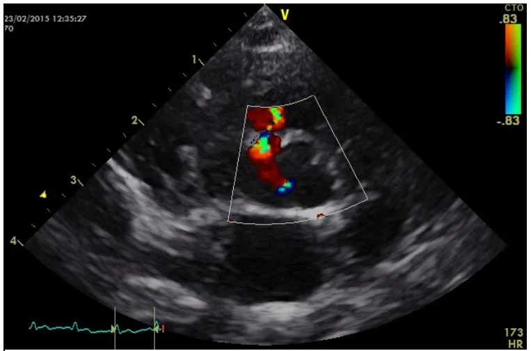
Colour doppler image courtesy of Emily Dutton at Cheshire Cardiology
A. Power doppler is very similar to colour doppler as a Colour Flow doppler. It too creates an overlay of flow in the colour window. Power doppler however measures amplitude rather than velocity and direction. As such you typically get a colour gradient such as dark to light orange showing increasing amplitude of returning signal. Due to the lack of direction information it is typically used for non-cardiac applications. It is more sensitive than colour doppler which is good for small and low velocity vessels. But this makes it more prone to flash artefact. This is where tissue movement is picked up filling the colour window with colour from movement such as breathing or panting.
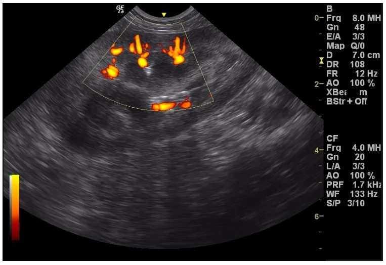
IMV Imaging image of power doppler
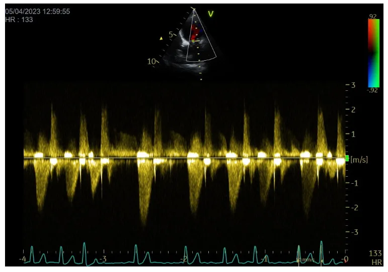
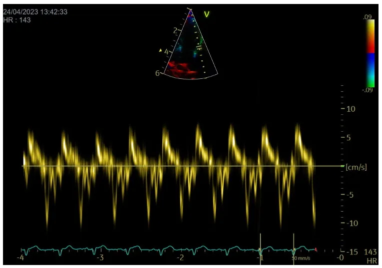
A. Pulsed wave doppler is a form of spectral doppler. It is used most commonly in cardiac applications but occasionally in abdominal work. It works by sending pulsed samples of sound into the patient as the name suggest. Pulsed wave doppler is depth specific. You get a sample volume you can position on the B-mode image. Which is where it will measure the velocity from.
This allows you to quantify the velocity of the blood in a specific region. Just like colour doppler (colour doppler being made up of many pulsed wave samples). There is a Nyquist limit beyond which aliasing will occur. This is a limitation of pulsed wave doppler. Particularly for fast jets in the heart, as you may not be able to quantify them with pulsed wave alone. Different machines will have different limits which is determined by the pulse repetition frequency the machine can achieve as well as the frequency it is emitting.

Pulsed wave doppler image courtesy of Emily Dutton at Cheshire Cardiology
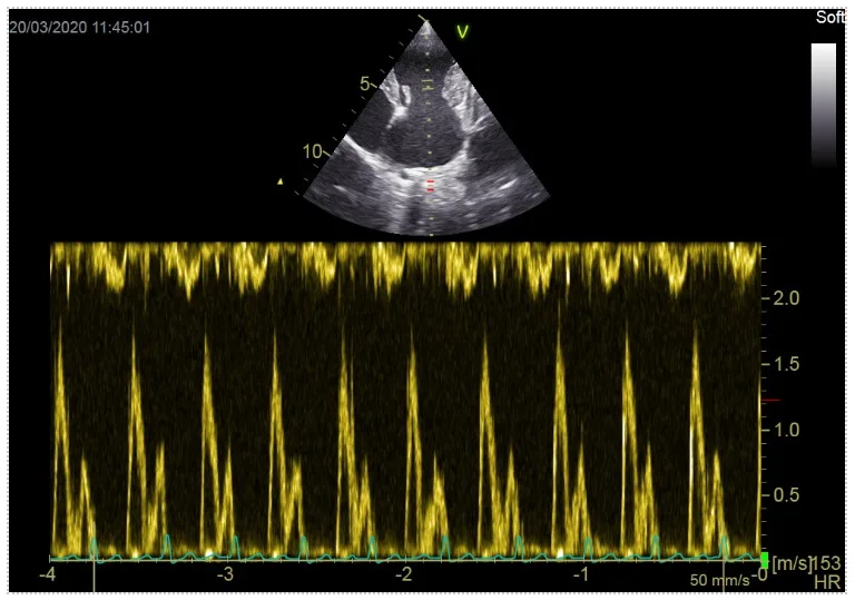
A. Continuous wave doppler is another form of spectral doppler. Unlike pulsed wave and again as the name suggests in continuous wave doppler sound is constantly emitted during the imaging process. This overcomes the limitations in pulsed wave of having to send and wait for returning pulses before sending the next one. This allows continuous wave doppler to measure a much higher velocity than pulsed wave doppler but with the trade off that we can no longer determine from where on the line the velocity was measured from. An assessment ahead of time with colour doppler should mean you know where you are expecting the highest velocity to originate from.
A. Tissue doppler really has to forms, Tissue Velocity Imaging, which is like colour doppler but for myocardium instead of blood, and Tissue Doppler Imaging which is like pulsed wave doppler but optimised for myocardium rather than blood. This allows you to make assessments of the motion of the myocardium. This is typically only used by cardiologists.
A. Aliasing is where the velocity you are exceeding exceed the scale the machine has been set to. When this happens the machine still measures the velocity but it mistakes it for a something going in the wrong direction. For example if you were using colour doppler and trying to assess a 100cm/s flow coming towards you (Red) with the scale set to 80cm/s the machine would interpret that as a 20cm/s flow going away from you (Blue). Being able to set you scale high enough to avoid this is important and can be a limitation of less expensive machines and higher frequency probes.
The deeper the structure you are trying to assess is the lower the maximum scale you can set will be in colour and pulsed wave doppler as the machine has to wait longer between pulses. For this reason bigger dogs can be more challenging. This same effect can be seen when filming a car wheel. If you have seen a video of a car wheel that seems to be going the right way but as the car speeds up it seems to start going the wrong way that is aliasing. Our pulses of sound are just like the frames of a video camera and we get the same effect in ultrasound.
A. First remember that colour doppler is directional. The machine measures things best that are moving directly towards and away from the probe. If you are trying to measure something perpendicular to the probe then it will not be picked up as a well. Try and reposition the probe to get better alignment if you can. Next make sure your gain is set appropriately. Position the colour window where you would like it to be, take the probe off the patient, turn the gain up until you get some noise, then down until it just goes away. Finally check your scale is set appropriately. You will want a lower velocity in the abdomen and a higher velocity in the heart. Remember when in a long axis cardiac view we have poor alignment with the probe and direction of blood flow. Even though 100cm/s would be normal in the heart you may find setting the scale to 75-80cm/s gives a better result in this view. Remember to change it back to 100cm/s or as close to that as possible for other cardiac views with better alignment to avoid aliasing.
A. We at IMV Imaging pride ourselves on our exceptional level of support. If you have any questions. Please don’t hesitate to get in touch with your local account manager or our head office.