05.09.2023

One of the most important steps to set yourself up for ultrasound success is choosing the correct ultrasound probe. Like with anything else, you need to pick the right tool for the job to get the best results.
Each probe will have pros and cons. Usually, the most important factors to decide on are resolution, penetration and footprint size.
These are some questions to think about:
Q. Can you give me a quick guide to the different probe options?
A. Sure, the common choices of probes are:
You need to select the correct probe that provides the best resolution for the required depth. Having a selection of probes allows for greater flexibility with what you can see.

Q. Ok, so how does the linear probe work?
A. Linear array probes are designed for high frequency, high details therefore superficial imaging.
The crystals inside the transducer are aligned in a linear fashion, perpendicular to the imaging face.
The flat head produces sound waves which travel out the face in straight line.
This is why the image produced on the ultrasound machine is easily identified by the rectangular in shape.
Q. Are there different types of linear probes?
A. Yes, there are two main types of linear probes: wideband linear array probes and hockey stick linear array probes.
We cannot forget the linear rectal probes, these guys work hard for our farm and equine colleagues. The imaging principles remain the same, just these probes are generally inserted rectally for image acquisition.
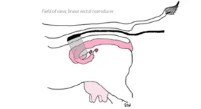
Field of view from a linear rectal probe. Image credit: Erika Wierman, DVM, 2022, Flat Linear vs. Curved Rectal Probe: How Do I Choose?, retrieved August 24, 2023, from: https://www.eimedical.com/blog/flat-linear-vs.-curved-rectal-probe-
Bovine Fetus – approx 71 days (left) and Equine Fetus – approx 26 days (right)
Q. When would I use which?
A. Wide band linear probe – Linear probes are good for superficial structures and for eye examinations.
One limitation is that they often have quite large footprints which may not be ideal.
Linear hockey stick probe – Hockey stick probes have an extremely small footprint making them a great choice for scanning exotic species, musculoskeletal structures in small animals and eyes.
Rectal linear probe – Although cows and horses are large animals, when scanning rectally, you are only a few centimetres away from reproduction and areas of the GI tract. For this reason, you can afford to use a high frequency transducer for much greater detail.
Q. When would you recommend reaching for a linear probe?
A. A general rule of thumb is that if you are going to ultrasound anything less than about 6cm deep, then use the linear probe. Anything deeper than 6cm you won’t be able to see much.
There are lots of scenarios where the linear probe is likely to be the best tool for the job, below are just some ideas to get you started:
Linear Rectal:
Anatomical location of structures can be reviewed.
Deeper abdominal organs can be examined, such as kidney, spleen and adrenal glands can be inspected.
Q. That’s a lot of information – can you give me a summary of linear probes?
These are great tools for:
To answer the question – Do I need one?
It is difficult to say, but there are lots of ways a linear probe can justify its place within your ultrasound toolbox if you get the opportunity to add one.